The MIN is led by Director Jeff Field[3], a physicist by training who also works closely with electrical engineering professor Randy Bartels to develop new imaging techniques and instrumentation[4]. “I love working on applying physics techniques and engineering tools to biological systems, and working at that interface,” Field said. “Every time I interact with someone who wants to solve a biology problem, I learn a little bit more – like what are the holes in imaging technology, and what are the things we’d like to do that don’t exist yet?”
The MIN operates under a fee-for-service model; users pay for time and training on the microscopes. Like all the foundational cores, the MIN offers Ph.D.-level skills and knowledge, through Field and a team of experts, to educate users and help them decide which instrument they need.
Deep roots
The MIN is managed through the Department of Biochemistry and Molecular Biology in the College of Natural Sciences. It has been a centrally operated campuswide resource for nearly 10 years, but its history stretches back further than that.
Jim Bamburg, emeritus professor of biochemistry and molecular biology, founded the MIN and serves on its board of directors.
“In 2008, when the first call for research infrastructure applications came out from the [Office of the Vice President for Research], we put forward a proposal to bring together many of the dispersed microscope units on campus under a single committee,” Bamburg said. “And we called this the MIN.” Since then, the model has saved researchers, colleges and departments hundreds of thousands of dollars in equipment purchases and service contracts. The MIN was officially named one of CSU’s Foundational Core Facilities in 2015.
“As faculty, all of us know where our money comes from,” Bamburg said. “Why shouldn’t we do the most we can for everyone, with the money we have, since people are investing in us and want the best to come from our results?”
What’s available?
As a result of its incremental history, MIN microscopes are scattered throughout different physical locations, including one at the Foothills Campus, as well as in the Molecular and Radiological Biosciences Building and the Anatomy/Zoology Building.
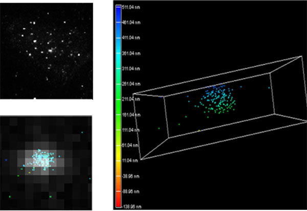
Among the offerings: two electron microscopes, three laser scanning confocal microscopes, a spinning disk confocal microscope, and two wide-field microscopes, as well as the superresolution TIRF-STORM that Nelson and Bailey use.
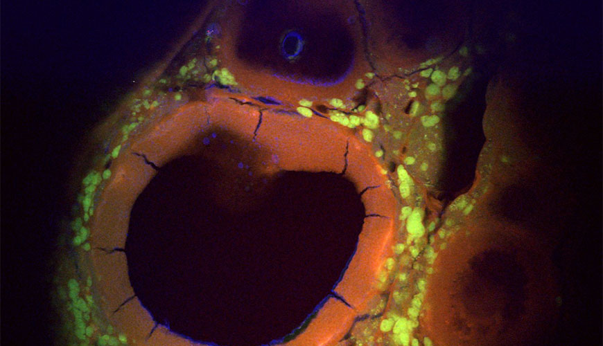
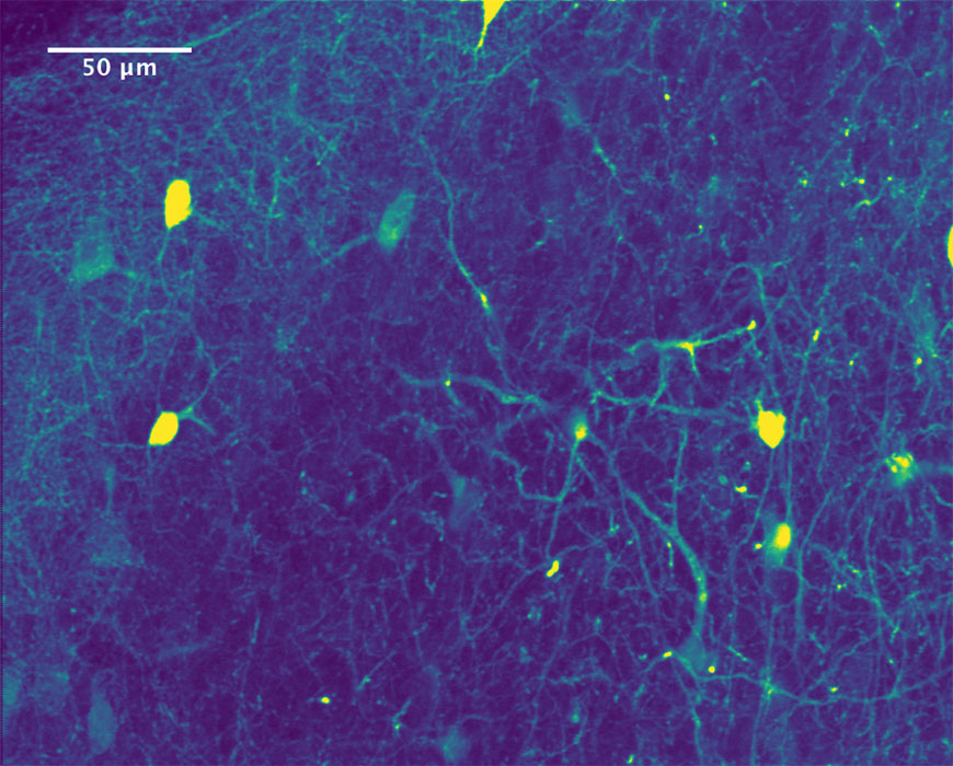
The TIRF-STORM and spinning disk microscopes detect fluorescence, and are especially designed for live-cell imaging.
Advances in fluorescence microscopy have allowed scientists to visualize things, while still alive and dynamic, that are just nanometers in length. Such microscopes are sensitive to fluorescent probes – glowing tags that bind only to certain proteins of interest – which have opened new doors for targeted, intracellular studies.
“These are things we used to have to extract and purify, and see how they behaved in test tubes,” Bamburg said. “Now, we can look at their behavior inside the cell. We can look at mitochondria, lysosomes, or even the nucleus of a cell.”
Electron microscopes, also available through the MIN, can go to much higher magnifications than fluorescence microscopes, but they’re not useful for live-cell imaging because they would destroy the samples. Electron microscopy requires vacuum chambers and fixed-tissue staining with heavy metals. These microscopes offer high-resolution, molecular-level imaging.
Customized solutions
In addition to providing access to commercial microscopes, the MIN can also provide pathways to customized instruments tailored to specific users’ needs. Field consults with potential MIN users to determine whether the microscopes in the core facility can provide what they need. In some cases, such as the telomere project, the right instrument for the job is already in place and ready for use.
Yet many researchers need data beyond what can be provided by a MIN microscope. In such cases, Field builds on over 12 years of experience designing lasers and microscopy systems for biological imaging to design new microscopes for the job.
In addition, the MIN funds a small number of pilot projects for faculty, in the form of facility access. A workshop series later this spring will cover some of these opportunities, as well as customized solutions that exist elsewhere on campus. Check the MIN website[5] for details.