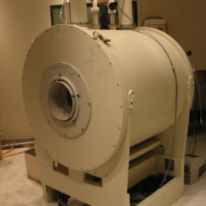
Using powerful magnets to produce a strong magnetic field, magnetic resonance imaging (MRI) produces three-dimensional, detailed images of objects or patients. Typically an MRI instrument is used for diagnoses and research within the brain, vascular networks, soft joint tissue and various organs, but the Rocky Mountain Magnetic Resonance team at Colorado State University has utilized the technology for other applications, as well.
RMMR, housed in the Department of Chemical and Biological Engineering, is a research facility specializing in the study of permeable materials. Experts there use MRI to, for instance, mathematically model processes within materials including underground geological media and the human body. Such studies form the basis for improved recovery of hydrocarbons, environmental remediation, as well as medical diagnoses.
Ted Watson, a professor and former head of the Department of Chemical and Biological Engineering, established and currently serves as the director of RMMR. The MRI instrument the lab houses, a Bruker Biospec Avance 24/30, has been involved in a number of studies from an array of disciplines.
From muscles to mice
Researchers from the College of Veterinary Medicine and Biomedical Sciences and the College of Health and Human Sciences are using the RMMR to study the architecture of leg muscles, and how that architecture changes over the lifetimes of Hartley guinea pigs that naturally develop osteoarthritis.
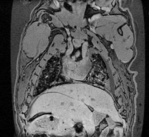
Watson’s team provided MRI images that showed successful repair of a surgically-induced defect underneath a kneecap for a project led by researchers in the CVMBS Orthopedic Research Center. Lab Animal Resources is also using the instrument to image live mice, looking at differential volumes of the cerebrum and cerebellum in mice exposed to different levels of cage enrichment.
Future applications
A potential future application of the MRI is osteoporosis detection. “We’d like to extend the technology to bone. Bone density is an indicator for osteoporosis, and permeability may be a better measure for detecting it,” said Benjamin Kohn, research associate at RMMR.
RMMR experts have developed a method to determine permeability distributions within materials like bone, which may provide a reliable method for identifying and assessing osteoporosis, a disease of increasing importance for the aging population.
Watson’s team hopes to make MRI technology available for other applications, and welcomes inquiries from the CSU community and beyond. RMMR is offering free imaging time for pilot studies to any CSU affiliated researchers for a limited time. To learn more about RMMR or MRI imaging, contact Benjamin Kohn.